
Introduction:
In This Article, We Tell You Details Info About the Atrioventricular Block of All First, Second, and Third Degrees.
Each Heartbeat starts with the heart’s pacemaker cells in the sinoatrial node, or sometimes just the SA node, in the right atrium.
The SA node transmits an electrical signal that propagates out through the walls of the heart and contracts both upper chambers, then moves through the atrioventricular (or AV) node where the signal stops for a split second, and then goes down into the lower chambers, where it moves down the bundle of His, into the left and right bundle branches, and each ventricles Purkinje fibers, causing them to contract as well.
So in a healthy heart, the upper chambers contract first, and then shortly after the lower chambers contract.
On an ECG, the atrial contraction is seen as a P wave, and the ventricular contraction is seen as the QRS complex. The interval of the start of the P wave to the origin of the QRS complex is called the PR interval plus is normally between 120 and 200 milliseconds or 3-5 tiny boxes on some graph paper that it’s usually printed out since each box is 40 milliseconds or 0.04 seconds.
Heart block describes a type of arrhythmia, or abnormal rhythm, that happens when the electrical signal gets held up and delayed or blocked entirely at some point along the conduction system. These blocks or delays usually happen because of some damage or fibrosis to the electrical conduction system, the pathways that conduct the electrical signal. Lev’s disease, or Lenegre-lev syndrome, is used to describe the large proportion of cases that are idiopathic and described as progressive cardiac conduction defects.
Causes:
- This means it’s not clear exactly what causes it, but over time, fibrosis, or scarring, develops in the conduction system which can delay or stop electrical conduction.
- This is usually a result of the aging process in the heart, and happens most usually in the elderly, although some genetic information has been recognized and can happen in younger people.
- Another large dimension of cases, though, is a result of ischemic heart disease, while the heart cells don’t get enough oxygen and can die off, as with a heart attack.
- This again leaves scar tissue that can block the electrical signal. In fact, it’s determined that about 20 percent of patients that have a heart attack go on to form a heart block.
- Finally, it’s deserving pointing out that the electrical conduction system is sort of like the electrical wiring in some walls of a home, so it makes sense that diseases of the heart muscle walls – cardiomyopathies, and inflammation of the heart muscle or myocarditis, can both cause heart block.
First, Second, and Third Degree Atrioventricular Block
An atrioventricular or AV block is used to describe when the signal is delayed or blocked when it’s trying to move from the atria to the ventricles.
First Degree Atrioventricular Block

- First-degree AV block when the signal is delayed but still makes it to the ventricles. This class has a PR interval greater than 200 milliseconds. Even though these signals are delayed, at the first-degree block, they still reach the ventricles. The first-degree block isn’t usually associated with any symptoms.
- Treatment or management of the first-degree block might involve identifying electrolyte imbalances or causes due to medications, although it usually doesn’t need further treatment.
Second Degree Atrioventricular Block
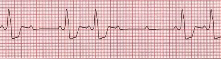
- Second-degree AV block can be split into two types. Type 1, called Mobitz 1 or sometimes Wenckebach, happens when the PR interval gets progressively longer with each beat until the P-wave is blocked completely.
- So maybe the first PR interval is 200 MS, then the next 260 MS, then 300 MS, and finally, the next one doesn’t make it to the ventricles, and you get what’s called a dropped beat.
- When a signal doesn’t do it to the ventricles from the atria, and if a long enough time passes by, let’s say about 2 seconds, and then ventricles pacemaker cells kick in, sort of as a fail-safe mechanism called a ventricular escape beat.
- Most people with this type don’t have symptoms, but occasionally patients feel lightheadedness, dizziness, and syncope.
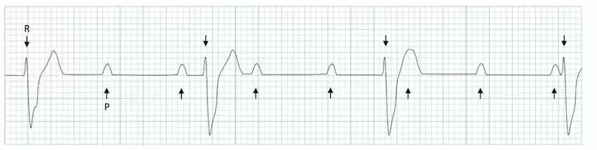
- Second-degree AV block Type 2, or Mobitz II, is similar to type 1 AV block in that you get irregular dropped beats, except this time they happen fairly randomly, so you don’t have this progressive lengthening of the PR interval.
- So if we get a more zoomed-out look at the ECG, some of these P waves have a QRS complex right after them, and some don’t and are followed by escape beats, what’s different here from Mobitz I is that each time it does conduct, the PR interval stays the same, as opposed to getting progressively longer.
- A lot of times a ratio for the overall number of beats conducted to not-conducted given, like 2:1 Mobitz II AV block, but again remember that the dropped beats happen randomly so we can’t predict exactly when the next beat will get dropped.
- Most people with Mobitz type II do feel symptoms from it like fatigue, dyspnea, chest pain, and syncope, though the severity can vary from patient to patient.
Third Degree Atrioventricular Block
- Third-degree, or complete heart block, describes when the signal is completely blocked when moving from the atria to the ventricles, every time. So in this case, even though the straight is going along at 60 bpm as if everything was normal, none of those signals make it down to the ventricles, and the ventricles struggle along with escape beats at prolonged rates, probably closer to 30 bpm.
- Patients with complete heart block are usually symptomatic, with symptoms ranging from syncope, confusion, dyspnea, and severe chest pain, and these patients are at risk of dying.
- When someone has AV block it’s important to find out the underlying cause and address it, for example, it could be an adverse effect from a medication or an infection.
- Regarding treating the AV block, typically a medication can be used to increase the heart rate like atropine. Alternatively, transcutaneous pacing might be used where the electrical pacing of the heart is done through electrodes placed on the skin.
- If the underlying cause is irreversible and the AV block is severe, patients ultimately might need a permanent pacemaker, which is implanted and continually monitors the patient’s rhythm, if it detects a delay, it will send an electrical impulse into the ventricles, causing them to contract
FAQs
An Atrioventricular Block is a condition where the electrical signals between the atria (upper chambers) and ventricles (lower chambers) of the heart are delayed or completely blocked. It is categorized into three degrees: first, second, and third degree.
In First Degree AV Block, there is a delay in the electrical conduction from the atria to the ventricles, but all signals eventually pass through. It is characterized by a prolonged PR interval on an electrocardiogram (ECG).
Second-degree AV Block may result in skipped heartbeats, dizziness, fainting, or chest discomfort. It is further classified into Mobitz Type I (Wenckebach) and Mobitz Type II, each with distinct ECG patterns.
In Third Degree AV Block (complete heart block), there is a complete blockage of electrical signals between the atria and ventricles. This leads to independent rhythms in the atria and ventricles, causing a slower and less effective heartbeat.
Symptoms may include fatigue, dizziness, shortness of breath, chest pain, and fainting. In severe cases, it can lead to heart failure or other serious cardiac complications.
Note:
So in This Post, Atrioventricular Block What other points can you think of/have experienced? Let me know in the comments.
If you found this helpful or feel free to share your experience if you can relate to these points and if you are comfortable share
For More Articles Related to Atrioventricular Block Stay Tuned To our Site: Health Daily Advice


